If your dentist has recommended dental X-rays and 3D scans, it is usually to see what cannot be checked properly with the naked eye. Dental X-rays help identify hidden decay, infections, bone loss, impacted teeth and problems under old fillings or crowns.
A 3D dental scan provides a more detailed view of the jawbone, nerves, sinuses and tooth roots for more complex treatment planning.
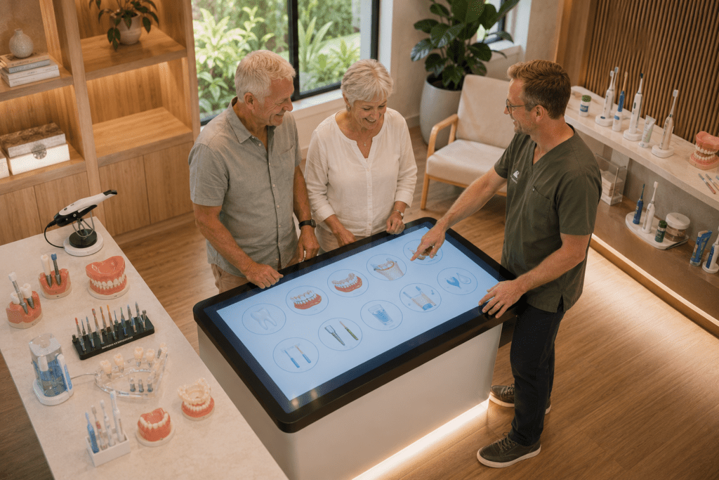
At Noosa Hinterland Dental in Pomona, we use low-dose digital imaging to support safer, more accurate dental care, including check-ups, wisdom teeth assessment, root canal treatment, implants, orthodontics and full-mouth planning.
Jump to section
- Why Do Dentists Use X-Rays And 3D Scans?
- Why Dentists Use Dental X-Rays And 3D Scans
- What Is A 3D Dental Scan And How Is It Different?
- When And Why Might Your Dentist Recommend Imaging?
- Are Dental X-Rays And 3D Scans Safe?
- What Happens During Common Dental X-Rays And 3D Scans?
- Ready To Talk About Dental X-Rays And 3D Scans?
- Frequently Asked Questions
Why Do Dentists Use X-Rays And 3D Scans?
Many people wonder why dentists recommend dental X-rays and 3D scans before treatment. A standard dental X-ray gives a flat two dimensional picture of teeth and nearby bone from one angle. A 3D dental scan, usually a cone beam CT scan, builds a detailed three dimensional model of teeth, jawbone, nerves and sinuses.
Regular dental X-rays use a very low radiation dose and work well for everyday checks, decay and gum assessment. A 3D scan uses a little more radiation, still in a low range, and is reserved for situations where extra detail protects important structures and guides complex care. At Noosa Hinterland Dental in Pomona, both options support safer treatment plans for families and adults across the Noosa Hinterland.
This article explains how dental X-rays and 3D scans work, when your dentist might suggest them, and how low dose digital technology keeps imaging gentle and reassuring. You will see how these tools help catch problems early and make treatment more accurate and comfortable.
If you have ever felt unsure about an X-ray or dental scan, the next sections aim to clear that up in plain language.
Key Takeaways
Before looking more closely at dental X-rays and 3D scans, it helps to see the main ideas in one place. These points explain why your dentist takes images and how Noosa Hinterland Dental keeps them safe and family friendly.
-
Why Seeing Below The Surface Matters
Many dental problems start between teeth or under old fillings where a mirror cannot reach. Dental imaging lets your dentist see these hidden areas long before pain or swelling appears. That means smaller treatments, fewer surprises and better long term tooth health. -
How Digital X-Rays And 3D Scans Support Safer Care
Clear images guide decisions about fillings, extractions, implants and braces so treatment is planned instead of guessed. At Noosa Hinterland Dental, digital systems give instant pictures that the dentist can zoom and measure on screen. This helps explain choices so you feel involved, not rushed. -
Radiation Dose And Dental X-Ray Safety
Modern digital dental X-rays use only a tiny fraction of the radiation from many medical scans. According to the Australian Radiation Protection and Nuclear Safety Agency (ARPANSA), a small dental X-ray is roughly similar to a day of natural background radiation. Careful settings keep exposure as low as reasonably achievable (the ALARA principle). -
When Imaging Is Especially Important – Implants, Wisdom Teeth, Root Canals
Extra detail from 3D scans helps protect nerves and sinuses during implant surgery and tricky wisdom tooth removal. Detailed views also guide root canal treatment when canals curve or split in unusual ways. These situations benefit most from three dimensional information. -
Why Noosa Hinterland Dental Uses Low Dose Modern Technology
The practice in Pomona invests in digital dental X-rays, cone beam CT and CEREC scanning so patients do not need to travel to Brisbane or Maroochydore. Low dose settings and careful selection of views match current Australian Dental Association guidance. This balanced approach suits children, anxious adults and people needing complex care.
Why Dentists Use Dental X-Rays And 3D Scans
Dentists use dental X-rays and 3D scans so they can see problems that eyes and mirrors simply cannot detect. At Noosa Hinterland Dental, this imaging supports early diagnosis, calmer visits and safer decisions for patients from Pomona, Cooroy, Cooran and Kin Kin.
Many dental diseases begin quietly inside teeth or under the gums. According to the Australian Institute of Health and Welfare (AIHW), around one quarter of Australian children aged five to ten have untreated tooth decay, much of it between back teeth. With digital dental X-rays, dentists can spot small cavities, bone changes and infections before they cause sleepless nights or broken teeth.
Imaging also helps with prevention. By comparing X-rays over time, your dentist can see if bone around your teeth is stable or slowly shrinking from gum disease. Research from the Australian Dental Association (ADA) shows that bone loss and decay are far easier to manage when found early. For Noosa Hinterland Dental, using images wisely means shorter, simpler appointments and fewer emergencies for local families.
“You can only treat what you can see. Dental imaging lets us find problems when they are still small and far easier to manage.”
— Clinical recommendation commonly supported by the Australian Dental Association
What Can Dental X-Rays Detect That A Visual Exam Can’t?
Dental X-rays reveal decay, infections and bone changes that are hidden beneath the surface, even when teeth look fine in the mirror. The main dental X-ray types used in Australia fall into two groups:
-
Intraoral views – bitewing and periapical images with a small sensor inside the mouth
-
Extraoral views – such as a panoramic dental X-ray, taken by a machine that moves around the head
Bitewing X-rays are common during check ups for children and adults in the Sunshine Coast hinterland. They show:
-
The tops of the back teeth
-
The bone level between them
This helps find:
-
Decay between teeth
-
Early signs of gum disease
Periapical X-rays show a whole tooth from crown to root tip and are invaluable when you have:
-
Toothache
-
Deep decay
-
A suspected abscess
-
Cracks or trauma affecting the root
Occlusal views and panoramic X-rays give a wider picture of the jaws. They help:
-
Find unerupted or extra teeth in children
-
Map wisdom tooth positions
-
Spot larger cysts or sinus issues
At Noosa Hinterland Dental, digital dental X-rays appear on screen within seconds, so the dentist can zoom, adjust contrast and explain what they see in everyday language. Imaging intervals are spaced according to your age, decay history and gum health instead of using a one size fits all schedule.
Conditions that may be missed without diagnostic imaging include:
-
Small cavities between teeth under tight contacts
-
Decay creeping under old fillings, crowns or bridges
-
Infections at the root tips before pain starts
-
Early bone loss from gum disease
-
Cysts or benign growths inside the jawbone
-
Impacted or missing teeth hidden in the jaw
What Is A 3D Dental Scan And How Is It Different?
A 3D dental scan is an advanced type of imaging that shows your teeth, jaws and nearby structures in three dimensions rather than as a flat picture. The most common form in dental practice is a cone beam CT (CBCT) scan.
During a CBCT dental scan, a cone shaped X-ray beam and a rotating scanner collect data from many angles around your head. Computer software then builds a three dimensional model of your upper and lower jaws, teeth roots, nerve canals and sinus spaces. According to RadiologyInfo, dental CBCT uses far less radiation than a full medical CT of the same area while giving richer detail than any standard dental radiograph.
Noosa Hinterland Dental also uses digital intraoral scanners, which create a 3D surface model of your teeth and gums without any radiation at all. These mouth scans use a small camera wand to record thousands of images every second and combine them into a precise virtual model. That model helps the team design CEREC same day crowns, bridges and clear aligners that fit comfortably and look natural.
3D Dental Imaging Vs Standard X-Rays: A Simple Comparison
3D dental imaging and standard X-rays both assist diagnosis, but they serve different roles. Two dimensional images are fast, low dose and ideal for routine checks. Three dimensional CBCT scans offer richer detail for higher risk procedures such as implants or complex extractions.
Standard Dental X-Rays Vs 3D CBCT Scans
| Feature | Standard Digital Dental X-Rays | 3D CBCT Dental Scan |
|---|---|---|
| What It Shows | Flat two dimensional view of teeth and nearby bone | Full three dimensional view of jaws, teeth roots, nerves and sinuses |
| Common Uses | Check ups, decay detection, gum disease review, emergency toothache | Implant planning, wisdom teeth near nerves, complex root canals, jaw joint and airway assessment |
| Radiation Dose | Very low, similar to a day or two of natural background in Australia (ARPANSA) | Higher than a small X-ray, but often still lower than a medical CT of the same region |
| Comfort And Time | Seconds per image while seated in the chair | Around half a minute standing or seated in an open scanner |
| How Often | At intervals based on decay and gum risk, often every one to two years for low risk adults | Only when extra detail will clearly change safety or treatment planning |
For example, before placing a dental implant in the front of the mouth, a CBCT dental scan shows exactly how thick the bone is and where nerves and sinus cavities sit. When planning a tricky wisdom tooth extraction in a Cooroy teenager, the 3D view helps the dentist judge how close the roots lie to the inferior dental nerve. In root canal treatment, CBCT sometimes reveals extra canals that standard films hide, which reduces the chance of ongoing infection.
When And Why Might Your Dentist Recommend Imaging?
Dentists recommend imaging at moments when extra information will protect your health, guide treatment or monitor changes over time. At Noosa Hinterland Dental, this applies from a child’s first bitewing X-ray to a CBCT scan for full mouth rehabilitation.
For families in Pomona, Cooran and Kin Kin, bitewing and panoramic X-rays help track how baby teeth fall out and adult teeth appear. These views can show:
-
Missing or extra teeth
-
Crowding
-
Impacted canines
-
Early jaw growth concerns
According to AIHW, early interceptive care often lowers future orthodontic complexity, so timely imaging matters.
Adults across the Noosa Hinterland may be offered dental imaging before:
-
Crowns and veneers
-
Invisalign style orthodontic treatment
-
Gum treatment for periodontal disease
-
A dental implant
-
Removal of wisdom teeth
-
Full mouth rehabilitation or complex restorative work
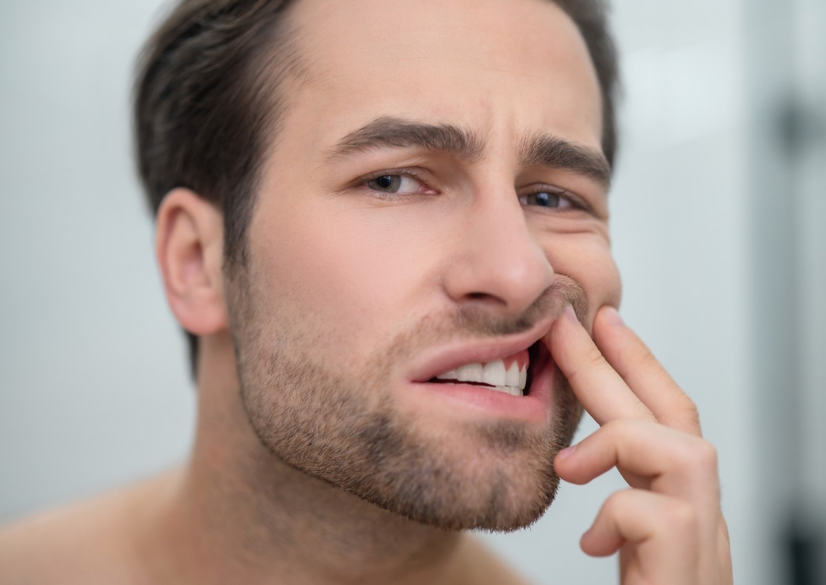
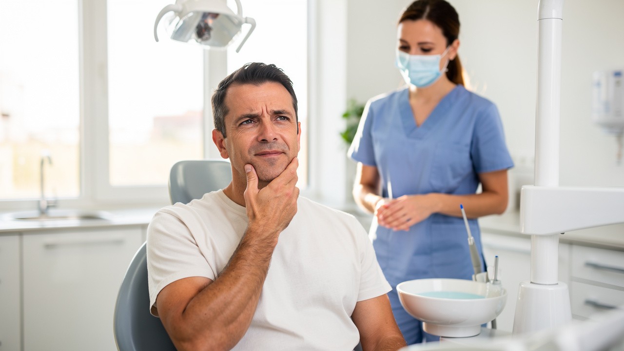
A periapical X-ray checks whether a heavily filled tooth is suitable for a crown, while a panoramic dental X-ray reviews wisdom teeth and jaw joints. During emergencies, such as sudden toothache or a sporting injury on the footy oval, fast X-rays help the dentist see if the cause is deep decay, a root fracture or an abscess.
Common situations where imaging may be recommended include:
-
Dental implants – CBCT for bone mapping and nerve location
-
Wisdom teeth – panoramic X-ray or CBCT to check angulation and nerve proximity
-
Root canal treatment – periapical X-rays and sometimes CBCT to locate all canals
-
Orthodontics – cephalometric and panoramic X-rays, plus 3D scans for aligner planning
-
Gum disease – bitewings and periapicals to monitor bone support
-
Hidden decay – bitewings to detect decay between teeth or under restorations
How Dental Imaging Supports Safer Treatment Planning
Dental imaging supports safer treatment planning by turning guesswork into measured decisions. For dental implants, a 3D dental scan lets the dentist at Noosa Hinterland Dental assess bone height, width and density in millimetres. The CBCT data combines with Implant Stability Quotient (ISQ) testing after placement so the team knows when an implant has integrated strongly enough for a crown.
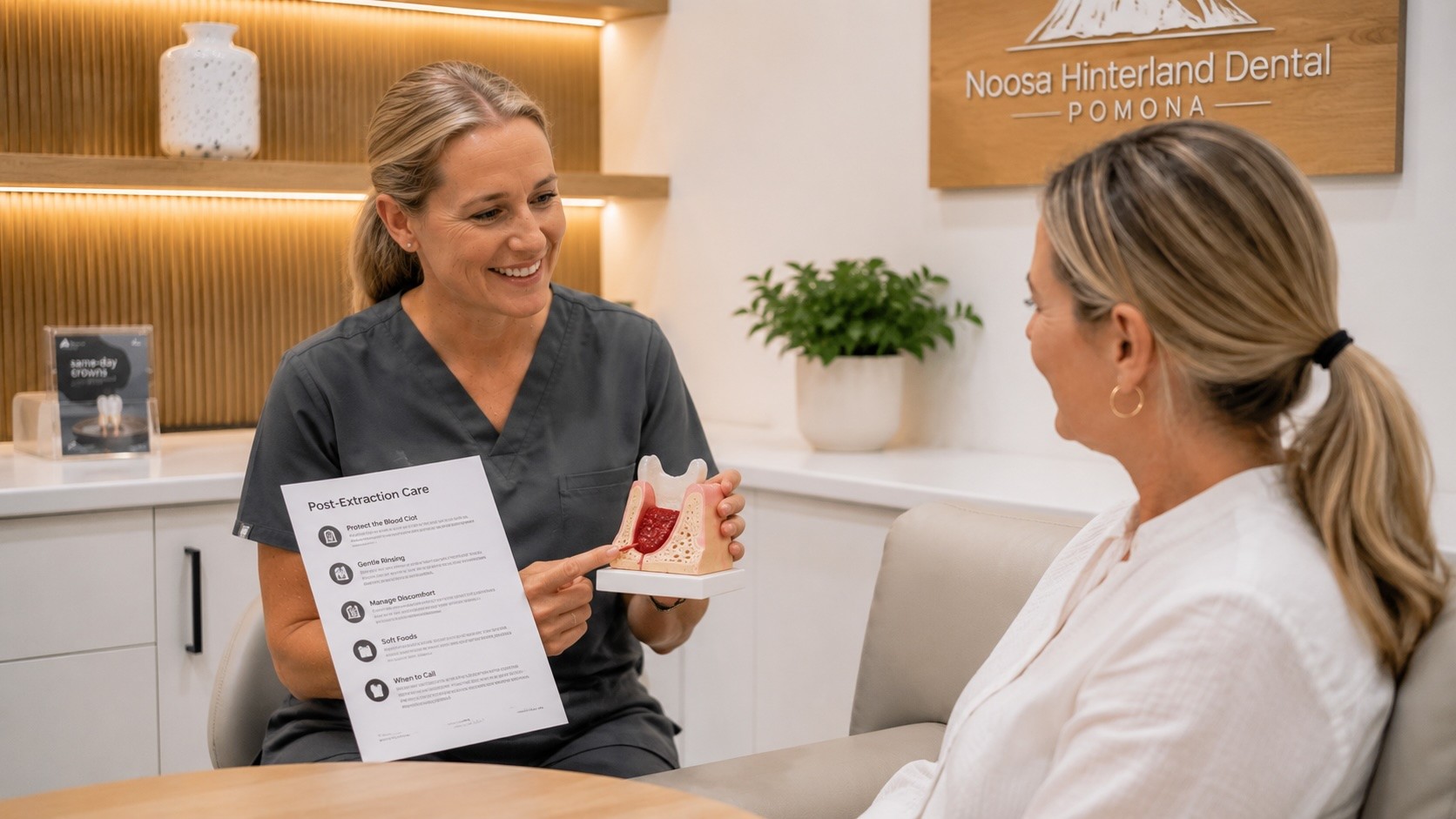
With wisdom teeth or other complex extractions, panoramic and CBCT images map root angles and their position near nerves and sinus floors. This helps decide whether a case can be managed comfortably in Pomona or is better referred to an oral surgeon in the wider Sunshine Coast. For root canal therapy, periapical X-rays taken before, during and after treatment check that the full length of each canal has been cleaned and sealed.
Orthodontic treatment, including Invisalign style clear aligners, relies on X-rays and 3D scans to confirm that bone support and root positions suit planned movements. In more extensive restorative work, such as full mouth rehabilitation for worn or missing teeth, imaging allows the dentist to design a stable bite that shares forces evenly. For anxious patients, clear pictures also help break treatment into gentle stages, which can be combined with sedation dentistry where needed.
Tip from the Noosa Hinterland Dental team:
“If you are unsure why an X-ray or scan is being suggested, ask your dentist, ‘What will this image show, and how will it change my treatment?’ A clear answer can be very reassuring.”
Are Dental X-Rays And 3D Scans Safe?
Dental X-rays and 3D scans are considered safe when used carefully, especially with modern digital equipment. The key point is that doses stay low and images are only taken when they provide a clear health benefit.
Digital dental X-rays at Noosa Hinterland Dental use sensors that need much less radiation than older film systems. ARPANSA explains that a typical small dental X-ray delivers only a tiny fraction of the radiation from many common medical scans. The X-ray beam is tightly focused on the mouth, and lead aprons or thyroid collars are used for children or when extra reassurance helps.
CBCT scans involve more radiation than a single bitewing, because they reveal a wider area in three dimensions. Even so, RadiologyInfo reports that a dental cone beam scan often uses far less dose than a standard medical CT of the same jaw region. For healthy adults, your dentist will only recommend 3D imaging if it changes diagnosis or makes a procedure safer, such as avoiding a nerve during implant placement.
Dental X-Ray Safety, Radiation Dose And Common Myths
Radiation is part of everyday life, from the sun, soil and some foods. According to ARPANSA, Australians receive several millisieverts of natural background radiation each year. A small digital bitewing X-ray is roughly equal to what you receive from simply living in Australia for a day or two, and a panoramic image still sits in a low range.
Safe dental X-rays for children use:
-
Child sized exposure settings
-
Careful positioning to avoid repeats
-
Only the views needed to monitor growth and decay
-
Protective shielding where appropriate
For pregnant patients, routine X-rays are usually delayed, but a localised image may still be recommended in an emergency, such as severe infection, because untreated dental sepsis carries its own risks. In these cases, the beam is aimed well away from the abdomen and shielding adds another layer of protection.
Common myths about dental imaging include:
-
“If my teeth don’t hurt, I don’t need X-rays.”
Research from the ADA shows that decay and bone loss often progress quietly without pain until they are advanced. -
“A CBCT scan is the same as a full body CT.”
In reality, a dental CBCT targets only the jaws and nearby structures, and usually uses a substantially lower dose than many medical CT protocols. -
“Digital X-rays are as strong as old film ones.”
Modern digital sensors are more sensitive, so the machine can run at much lower settings.
At Noosa Hinterland Dental, the team explains these points, invites questions and offers sedation options for very anxious patients who worry about any procedure.
What Happens During Common Dental X-Rays And 3D Scans?
Knowing what happens during dental X-rays and 3D scans can make the whole process feel far less intimidating. Most images are completed in seconds to a minute, with little or no discomfort.
For intraoral X-rays such as bitewings or periapicals, you usually sit in the dental chair at Noosa Hinterland Dental:
-
A small sensor rests gently inside your mouth.
-
You close your teeth together to hold it still.
-
The X-ray arm moves next to your cheek while you stay still for a moment.
-
The image appears almost instantly on the computer screen for review.
A panoramic dental X-ray, often called an OPG, involves standing or sitting in a special machine:
-
Your chin rests on a support and you may bite on a small guide.
-
The unit slowly rotates around your head without touching it.
A CBCT 3D dental scan feels similar, although the scanner may rotate for slightly longer to gather extra information.
Digital intraoral scanning for CEREC crowns or aligners uses a slim camera wand passed over the teeth to build a 3D model, without any radiation at all.
What To Expect At Noosa Hinterland Dental In Pomona
At Noosa Hinterland Dental in Pomona, every step of imaging is designed to feel calm and clear. The team explains which pictures they recommend, whether that is a couple of bitewing X-rays, a panoramic view or a CBCT dental scan for implants or wisdom teeth. Children are shown the small sensors and the larger machines first, so the experience feels familiar rather than strange.
During an intraoral X-ray, the dentist or assistant places child sized or adult sized sensors gently and checks your comfort before taking the image. For panoramic and CBCT scans, you stand or sit in an open style unit, supported at the chin and forehead so you do not have to hold your head stiffly. The rotation is smooth and takes less than a minute, and you can breathe and swallow normally.
Digital files from every dental radiograph and 3D dental imaging session are stored securely and can be compared over time. This helps monitor changes in teeth, gums and jawbone, which is handy for people with gum disease, implants or long term restorative work. For anxious patients from Cooroy, Cooran, Kin Kin and Noosaville, comfort measures such as blankets, reclining chairs, ceiling mounted screens and noise cancelling headphones are available. Sedation dentistry can be arranged for those who find even short procedures very stressful, so a needed dental scan does not have to feel overwhelming.
Ready To Talk About Dental X-Rays And 3D Scans?
Dental X-rays and 3D scans give your dentist a clear view of what is happening under the surface, which helps catch problems early and plan safer treatment. With low dose digital systems and careful selection of views, these tests sit in a very low radiation range for most people.
Noosa Hinterland Dental combines modern imaging with a gentle, family centred approach for residents across Pomona, Cooroy and the wider Sunshine Coast hinterland. Whether you are thinking about implants, Invisalign, cosmetic work or simply want reliable care for your children, the team can explain exactly why a particular image is suggested and how it will influence your treatment plan.
Conclusion
When used thoughtfully, dental X-rays and 3D scans become quiet helpers that protect your smile rather than something to fear. Two dimensional images suit day to day checks, while three dimensional CBCT scans support complex care such as implants and tricky wisdom teeth. Low dose digital technology, careful timing and clear explanations all reduce worry. If you have questions about imaging or radiation, talking them through with the team at Noosa Hinterland Dental can bring welcome peace of mind.
Frequently Asked Questions
Question: How Often Do I Really Need Dental X-Rays?
Most people need dental X-rays every one to two years, but timing varies. Low risk adults with healthy mouths may need images less often, while children or adults with a history of decay or gum disease may benefit from more regular checks. At Noosa Hinterland Dental, X-rays are never taken routinely without a clear reason.
Question: Is A 3D CBCT Dental Scan Necessary For Every Implant?
A 3D CBCT dental scan is recommended for most implant plans because it shows bone volume and nerve or sinus positions very clearly. In simpler cases with plenty of bone, your dentist might rely on standard X-rays instead. The team at Noosa Hinterland Dental will explain which option suits your mouth and why.
Question: Are Dental X-Rays Safe For Children In The Noosa Hinterland?
Yes, dental X-rays are considered safe for children when used carefully with child sized settings and shielding. They help pick up decay between teeth and guide growth and orthodontic decisions. For families around Pomona and Cooroy, the small radiation dose is balanced against the benefit of finding problems long before pain starts.
Question: What If I’m Pregnant And Have A Dental Emergency?
If you are pregnant and in pain, a localised dental X-ray may still be recommended to treat serious infection or trauma. The beam is aimed at the mouth, not the abdomen, and a lead apron adds extra protection. Routine images are usually delayed, and your dentist will discuss options with you before any exposure.
Question: Can I Refuse Dental X-Rays Or 3D Scans?
Yes, you can decline any test, including dental X-rays and 3D scans. Your dentist will explain how refusing images may limit diagnosis or make treatment less predictable. At Noosa Hinterland Dental, the team can discuss alternatives, timing and the risks of waiting so you can make an informed choice.
Question: Will My Private Health Insurance Help Cover Dental Imaging Costs?
Many extras policies contribute to the cost of dental X-rays and some 3D scans, although cover varies between funds. The reception team at Noosa Hinterland Dental can provide item numbers and estimates so you can check your policy. Most routine digital dental X-rays are relatively affordable and are only recommended when they genuinely add value to your care.